
Usage in organs
Breast biopsy marking
Prove the correct location of the biopsy
The majority of the suspicious lesions detected by screening mammography are small nonpalpable lesions, i.e. mass lesions or calcifications. Biopsy are performed to differentiate between benign and malignant lesions and decide if surgical removal is needed.
It is helpful to mark these lesions with a marker (clip) which can be implanted during the initial biopsy procedure or in a second step. Clip-marking is done in order to prove the correct location of the biopsy and to guide the surgeon for the excision.
Clip-marking can also be useful for the follow up of suspicious lesions with benign histology.
Breast biopsy marker brochure
Mammogram and MRI showing Gold Anchor implanted in breast to mark a biopsy site
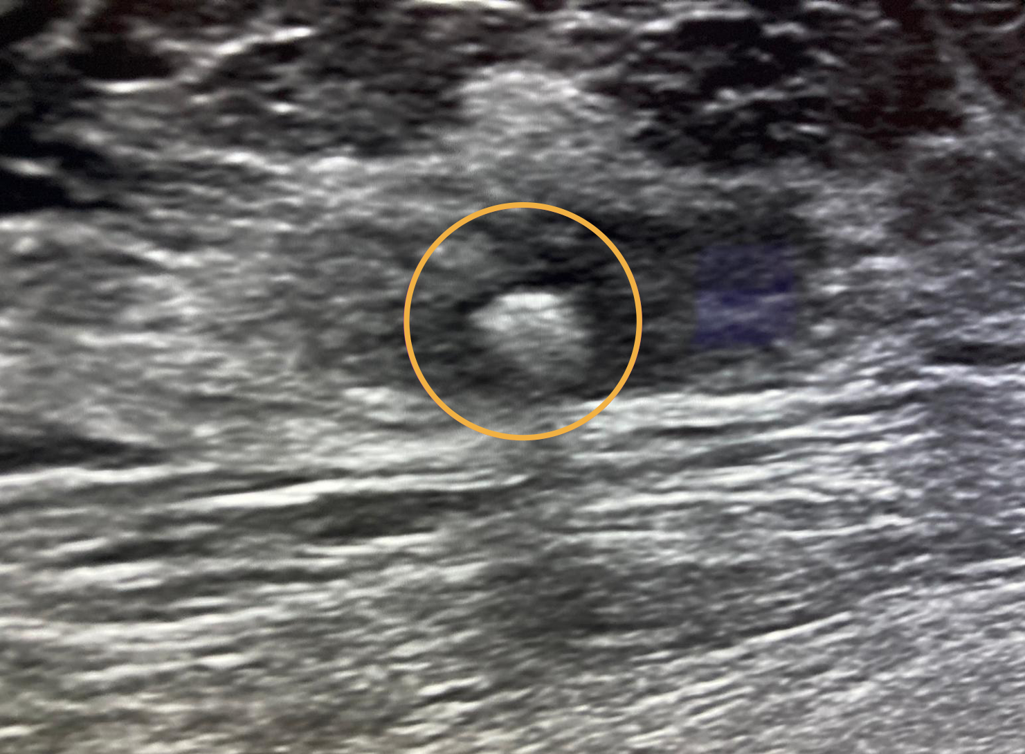
Ultrasound image showing Gold Anchor implanted in breast to mark a biopsy site.
Define the area of breast tissue which must be removed
Before surgery, these clips are usually used for guide wire placement. Exact positioning of the clips within or right next to the lesion helps to reduce the size of tissue that should be surgically removed. For some tumors neoadjuvant treatment (i.e. chemotherapy and/or hormone treatment before surgery) is used for downsizing the tumor prior to surgery. Clip-marking in these cases is important in order to define the area of breast tissue which must be removed.
Reduce implantation time
- Gold Anchor is ideal for breast biopsy marking with its unique and patented design.
- The Gold Anchor markers are pre-loaded in industry leading thin 20G needles.
- When deployed, Gold Anchor expands outside the needle and attaches immediately into the tissue.
- Gold Anchor can be implanted percutaneously, guided by stereotactic mammography, ultrasound or CT.
