
Exceptional visibility on MRI
Great visibility

Thin marker in
unique material
Gold Anchor has excellent visibility on imaging modalities such as kV, CT, CBCT, MRI and Ultrasound. The marker is made of an alloy of pure gold mixed with a small portion of pure iron for improved MRI visibility (Int. patents).
Gold Anchor comes in four different sizes and can be implanted with either a line or ball shape. This flexibility allows for controlled image artefacts on various imaging modalities. The marker diameter is only 0.28 or 0.4 mm, which improves the surface-to-volume ratio.
Benefits
- Clearly visible on kV, CT, CBCT, Ultrasound and MRI
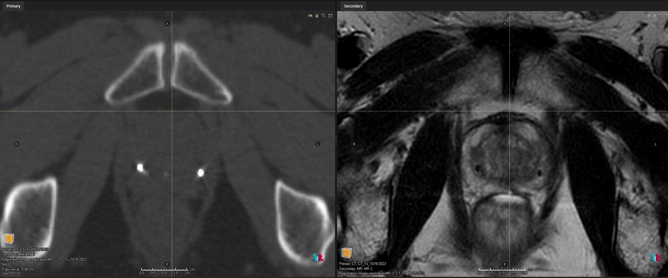
- Easy registration of CT and MR images
- Enables Intrafraction Motion Management
- Low dose perturbation for Particle Therapy
- Reduced CT/CBCT artifacts
- Ideal for MR-only workflow
- Great for biopsy marking and surgical guidance
Gold Anchor MR+
The Gold Anchor MR+ product line is the latest addition to the Gold Anchor product portfolio. The MR+ product range has a unique and patented marker material, an alloy of 98.5% pure gold and 1.5% pure iron. (Earlier product version of Gold Anchor is made of 99.5% iron mixed with 0.5% pure iron, please note that the Gold Anchor MR+ product line is not available for sale in all Gold Anchor markets).
The MR+ marker material provides great MRI visibility, especially on T2-weighted images, which typically are used for prostate delineation. This helps when fusing CT and MR images and when moving to MR-only workflows.

CBCT of 0.28×10 mm Gold Anchor MR+ implanted with ball shape in prostate.

T2-weighted MRI of 0.28×10 mm Gold Anchor MR+ implanted with ball shape in prostate.

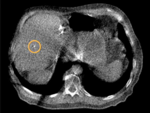
CT of 0.4×10 mm Gold Anchor MR+ implanted with ball shape in prostate.

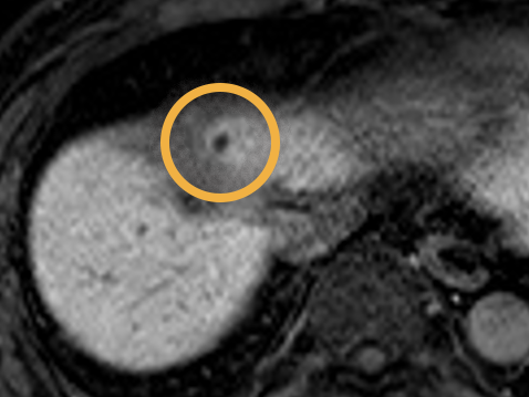
T2-weighted MRI of 0.4×10 mm Gold Anchor MR+ implanted with ball shape in prostate.

Line shaped 0.4 mm diameter Gold Anchor MR+ marker in prostate visualized on T2-weighted MR- sequence. Image courtesy of Dr. Marcio Fagundes at Miami Cancer Institute.

Line shaped 0.28 mm diameter Gold Anchor MR+ marker in prostate visualized on T1-weighted MR-sequence. Image courtesy of Gjøvik Hospital, Norway
Easily register CT and MR images
Ball shaped Gold Anchor MR+ markers can typically be visualized directly on the T2-weighted MR sequences used for prostate delineation. This is very helpful when fusing CT and MR-images and may eliminate the need for an additional MR “marker sequence” and thereby enable a more precise fiducial-based image fusion. Read more here.

Minimize artifacts and dose perturbation
For minimal CT artifact the Gold Anchor markers should be implanted with a line shape. This is ideal for particle therapy since it minimizes dose perturbation. The ultrathin 0.28 mm diameter Gold Anchor MR+ marker cause very low CT artifact while still being visible on T1-weighted MR-sequence as shown in the prostate image.
Image courtesy of Gjøvik Hospital, Norway
More MR images and settings Spatial Accuracy and Visibility on MRIEnables Intrafraction Motion Management
Clearly visible on kV-imaging
Gold Anchor has been designed for use with kV imaging. Using Gold Anchor with kV-based Image Guided Radiotherapy enables a more accurate and reliable treatment process. Trackable with Accuray Synchrony® (CyberKnife® and Radixact®), Brainlab ExacTrac Dynamic® and with Varian Truebeam® Auto Beam Hold to detect and adjust for intrafraction motion.
The image shows a ball shaped 0.4×20 mm Gold Anchor used for positioning of a liver SBRT patient. The MVCT image was acquired with the TomoTherapy® Hi Art® treatment system immediately prior the treatment.
Visible on MVCT
Gold Anchor markers are visible on MVCT images across various imaging systems. Typically, the largest Gold Anchor marker (0.4×20 mm), implanted in a ball-shape configuration, is recommended for optimal MVCT visibility. However, advancements in imaging technology, such as the Accuray Radixact system with its CTrue™ IR (Iterative Reconstruction) algorithm, allow for the visualization of smaller Gold Anchor markers as well. For additional details, please refer to the section on enabling technologies for Accuray TomoTherapy and Radixact MVCT.

Image showing a ball-shaped 0.28×10 mm Gold Anchor marker implanted in prostate imaged with Accuray Radixact. Image courtesy of Capital Regional Cancer Center, Tallahassee, FL, USA.

Images courtesy of Dr. Hiroshi Doi, Mr. Shogo Harui and Dr. Yoshio Hishikawa at the Meiwa Cancer Clinic, Japan
Images courtesy of Dr. Hiroshi Doi, Mr. Shogo Harui and Dr. Yoshio Hishikawa at the Meiwa Cancer Clinic, Japan