
A Great Fiducial Marker
CT/MRI fusion
Great visibility in CT and MR images
Gold Anchor MR+ offers great visibility in CT and MR images allowing for reliable and efficient MR/CT fusion.
Prior to receiving radiation therapy, patients typically undergo computed tomography (CT) simulation. CT images enable accurate dose calculation in treatment planning systems. However, due to the superior soft tissue contrast, many patients also undergo Magnetic Resonance Imaging (MRI). Target volume delineation is then typically performed on T2-weighted MRI.
Implanted fiducial markers can make the CT/MRI fusion more reliable and efficient. However, traditional gold markers are often not seen well on T2-weighted MRI. Therefore, many radiotherapy centers use an additional “marker sequence” (e.g. T1-weighted) to be able to identify such markers on MRI. This is not ideal since a shift in patient/target between the different MR images can cause a systematic setup error in later image-guided radiotherapy (IGRT).
Gold Anchor MR+ markers provide enhanced MRI visibility due to the unique and patented marker material (an alloy of 98.5% pure gold and 1.5% pure iron). This may eliminate the need for an additional MR “marker sequence” and thereby enable a more precise fiducial-based image fusion.
Images showing 0.4×10 mm Gold Anchor MR+ markers, implanted with a ball shape in prostate. Images courtesy of Centralsjukhuset Karlstad.
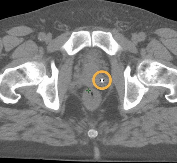
CT

T2-weighted MRI

“The Gold Anchor MR+ markers provide great visibility on MRI. When we shifted to 0.4×10 mm Gold Anchor MR+ markers, implanted with a ball shape in prostate, we found that we no longer need to capture T1-weighted MRI sequences. We can now reliably identify the markers on the T2-weighted sequences that we use for target delineation.”
Hans-Olov Rosenbrand
Medical Physicist, Centralsjukhuset Karlstad, Sweden